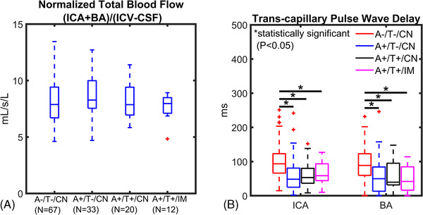
FIGURE 1.

Blood flow and transcapillary pulse wave delay from cardiac‐resolved 4D flow magnetic resonance imaging. Data are summarized for controls and amyloid/tau (AT) biomarker–confirmed groups (amyloid positive [A+], tau positive [T+], cognitively normal [CN], cognitively impaired [IM]). A, Cerebral blood flow (basilar [BA] + internal carotid artery [ICA]) was normalized to brain volume and was similar between groups. B, ICA and BA transcapillary pulse wave delay time was significantly longer in the control group (A–/T–/CN) compared to A+/T–/CN (ICA P < .001; BA P = .001), A+/T+/CN (ICA P < .001; BA P = .050) and (A+/T+/IM; ICA P = .003, BA P = .026) indicating a significantly faster transmission of the cardiac pulse wave in the intracranial vascular network of AT biomarker–confirmed groups
