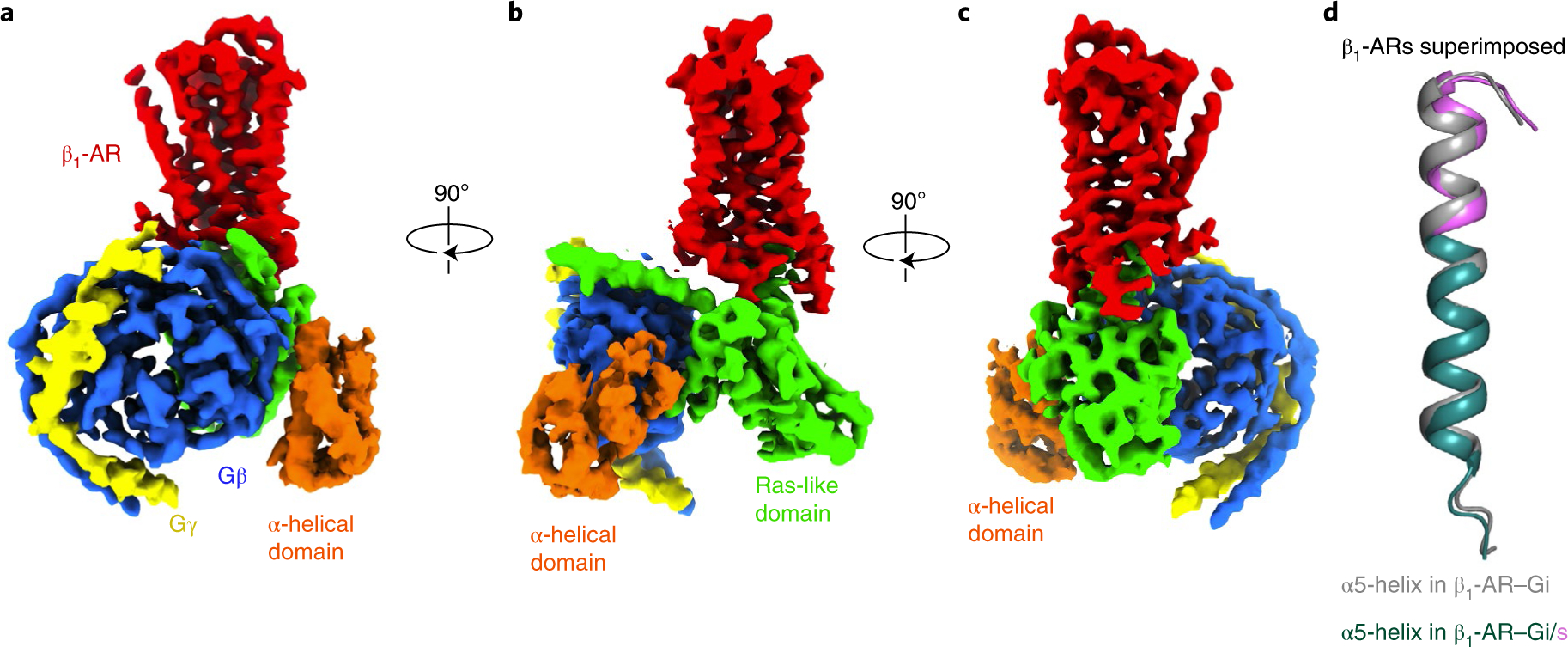
Fig. 6. Cryo-EM structure of the complex of isoproterenol-bound β1-AR and Gi/s.

a–c, Cryo-EM density maps, in three views (a, b and c), of the β1-AR-Gi/s complex colored by subunit (β1-AR in red, Gαi/s Ras-like domain in green, Gαi/s α-helical domain in orange, Gβ in blue, Gγ in yellow). d, Comparison of the α5-helices in the β1-AR–Gi complex (gray) and the β1-AR–Gi/s complex (in dark teal with the last 11 amino acid residues from Gαsin violet) when β1-ARs are superimposed.
