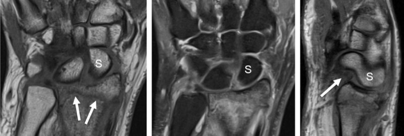
Figure 2.

Distal radius fracture with associated intrinsic scapholunate (SL) and extrinsic palmar radiocarpal ligament rupture (RSC, radioscaphocapitate) detected by MRI. T1 coronal slice (a) shows the fracture line (arrows) and the increased SL space. Proton-density fat saturated coronal slice (b) demonstrates high signal surrounding bone marrow edema and increased signal of the SL space. Sagittal proton-density slice (c) detects a dorsal avulsion of the distal radius and a torn palmar RSC ligament (arrow) with rotatory subluxation of the scaphoid (S).
