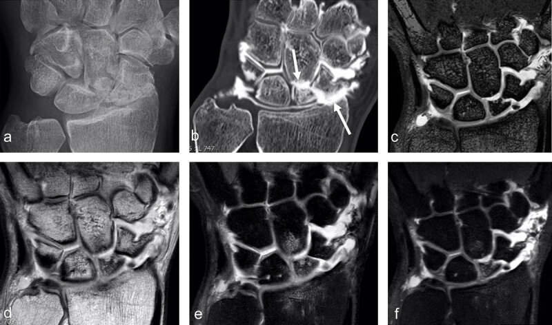
Figure 4.

SNAC 2 wrist after scaphoid fracture with nonunion in a 41-year-old man. Secondary cartilage degeneration is first seen in the radioscaphoid compartment (b, arrow pointing up) and later in the scaphocapitate midcarpal joint space (b, arrow pointing down) on conventional radiograph and much better on CT-arthrography (b). MRI well demonstrates the nonunion (on 3D DESS with 0.4mm sections) and increased signal in the proximal scaphoid pole on 2D (e) and 3D (f) proton-density fat-saturated images, corresponding to edema and indicating that the separated fragment might be unstable (and painful). Subchondral bone marrow edema is also detected in the proximal pole of the capitate under the cartilage loss and in the distal radius (e).
