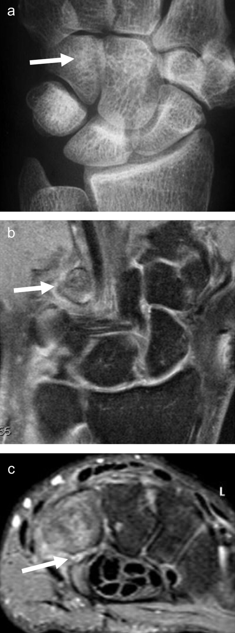
Figure 6.

Fracture of the hook of the hamatum, easily missed on conventional radiographs and on fat-saturated MR images. The AP X-ray (a, arrow) demonstrates the absence of the ring sign (normally present at the level of the hamulus) on the hamate bone. On palmar proton-density fat-saturated coronal image (b, arrow), the presence of BME within the hamulus may be obscured because of the close signal intensity of the surrounding muscles. The fracture line at the base of the hamulus could also be missed on the axial proton-density fat-saturated sequence (c, arrow).
