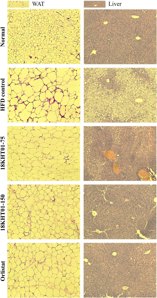
FIGURE 8.

Effect of 18KHT01 on white adipose tissue (WAT) and liver histology. Hematoxylin and eosin (H&E) staining was done to perform histological analysis. Pictures were captured at ×20 magnification under a light microscope. Size of adipocytes and crown-like structure (red eosin stain) were evaluated in WAT. Accumulation of lipid droplets (yellow droplets) and macrophage infiltration are evaluated in the liver. Lipid droplets are more noticeable in HFD control liver.
