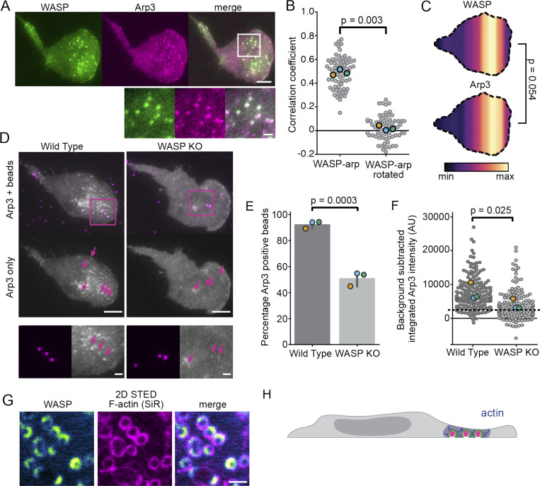
Figure 6.
WASP mediates actin polymerization at sites of membrane deformation. (A) Endogenous WASP colocalizes with TagRFP-T-Arp3. Scale bars are 5 μm and 1 μm in the inset. (B) Pearson correlation coefficients of WASP and Arp3 show significant colocalization (r = 0.49 ± 0.01) compared with the correlation coefficients between WASP and a 90° rotation of the Arp3 channel (0.02 ± 0.02). n = 68 7.5 × 7.5–μm regions of interest collected across three experiments. P = 0.003 by a paired two-tailed t test on replicate means, which are denoted throughout the figure by enlarged markers. (C) Spatial distributions of WASP and Arp3 puncta reveal similar organization of these proteins. n = 18 coexpressing cells collected across three experiments with nWASP = 559 and nArp3 = 417 total puncta. The mean relative puncta position is 0.64 ± 0.02 for WASP and 0.65 ± 0.02 for Arp3, with the cell rear defined as 0 and the cell front as 1. P = 0.054 by a paired two-tailed t test on the mean puncta position of each marker in each cell. (D) WASP KO cells fail to recruit appreciable amounts of the Arp2/3 complex to 100-nm bead-induced invaginations. Arrows denote the position of beads that are under the cell front. Below is a zoomed inset of the boxed region. All images are scaled equally. Scale bars are 5 μm and 1 μm in the inset. (E) Only about half of the beads (51.3 ± 3.1%) under the cell front in WASP KO cells are able to form Arp2/3 puncta (determined by the ability to fit a Gaussian to Arp3 signal at a bead) compared with 93.7 ± 1.7% of beads for WT cells. nWT = 172 beads and nKO = 130 beads collected across three experiments. P = 0.0003 by an unpaired two-tailed t test on replicate means. (F) Arp2/3 complex recruitment to bead-induced invaginations is significantly less in WASP KO cells than in WT cells. For each bead, the integrated intensity of background-subtracted Arp3 signal was calculated for the brightest Arp3 frame. The dashed line marks the approximate threshold needed to fit a Gaussian in panel E. Measurements below this line are close to background or noise. The mean intensity of Arp3 at beads is 7,696 ± 1,454 a.u. in WT cells and 4,020 ± 886 a.u. in WASP KO cells on the replicate level. nWT = 164 and nKO = 143 beads collected across three experiments. P = 0.025 by a paired t test on replicate means. (G) WASP and F-actin (reported with SiR-actin) colocalize at bead-induced membrane invaginations in the front half of the cell. Scale bar is 1 μm. (H) Updated model to reflect front-biased WASP-dependent actin polymerization. AU, arbitrary units; max, maximum; min, minimum.

