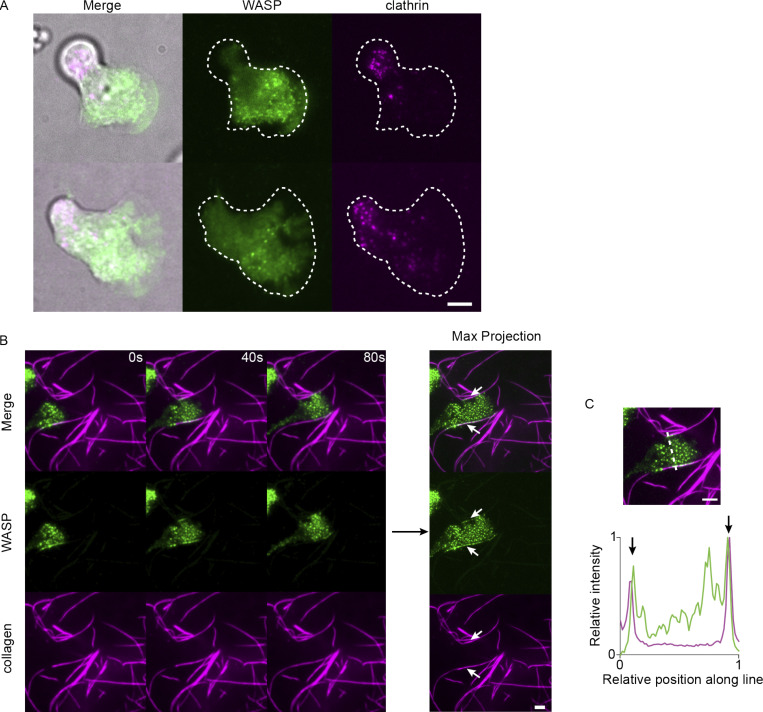
Figure S2.
WASP puncta formation in the absence of agarose overlay and on collagen fibers. (A) Endogenous WASP forms front-biased puncta and is spatially segregated from clathrin LCa in the absence of agarose overlay. (B) As a more physiological counterpart to bead-induced membrane invaginations, we imaged WASP in cells migrating across fluorescent collagen-coated coverslips and found that WASP is recruited to sites of cell contact with fibers. Right-hand side shows the maximum (max) intensity projection over time, which further highlights the enrichment of WASP to cell–fiber interfaces. Stretches of contact are marked with arrows. (C) Relative background-subtracted WASP (green) and collagen (magenta) signal along the dashed line drawn on the top image. Arrows denote WASP puncta formed at sites of cell–collagen contact. Scale bars are 5 μm. Imaged with TIRF microscopy.

