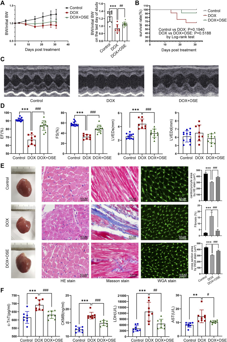
FIGURE 2.
NEU1 inhibitor improved DOX-induced cardiac dysfunction in rats. (A) The tendency of body weight change of rats in three groups (n = 12 per group). (B) The survival curve of rats in different groups. (C) Representative images of M-mode echocardiography in different rat groups at the endpoint of the study. (D) Statistical analyses of echocardiographic parameters including ejection fraction (EF), fractional shortening (FS), left ventricular end-systolic dimension (LVEDs), and left ventricular end-diastolic dimension (LVEDD) of rats in the three groups at the end of the study. (E) Representative images of hematoxylin and eosin (HE) staining, Masson’s trichrome staining, and wheat germ agglutinin (WGA) staining of heart tissues in the three different groups, which display the extent of cardiac atrophy, myocardial fibrosis, and cell sizes, respectively (scale bar = 50 μm). (F) Concentrations of cardiac injury markers including cardiac troponin T (cTnT), creatine kinase isoenzyme-MB (CK-MB), lactate dehydrogenase (LDH), and aspartate aminotransferase (AST) in plasma of rats in different groups. (**p < 0.01 vs. control group, ***p < 0.001 vs. control group. #p < 0.05 vs. DOX group, ##p < 0.01 vs. DOX group, ###p < 0.001 vs. DOX group).

