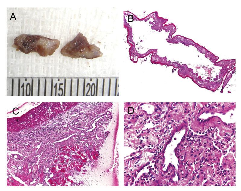
Figure 2.
Macroscopic and histopathological aspects of angina bullosa haemorrhagica. (A) The gross specimens showed irregular shapes and cut surfaces with whitish and brownish areas. (B) Oral mucosa lining epithelium showing areas of detached connective tissue and hemorrhage. (C) Area of ulceration and granulation tissue and (D) diffuse mixed inflammatory infiltrate. (B-D, H, E stain; original magnifications: B, 40×; C, ×200; D, ×400).

