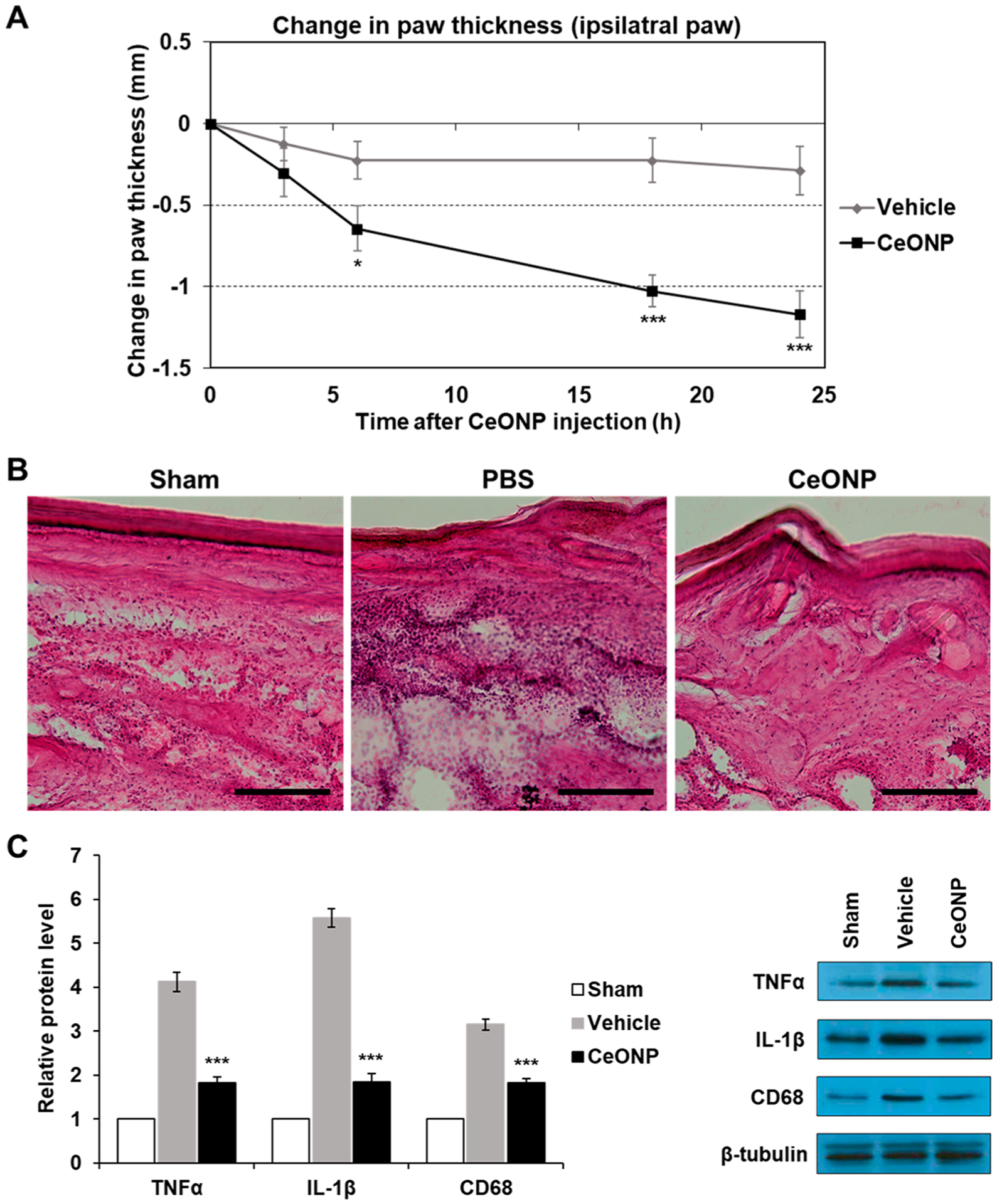
Figure 7.

In vivo edema reduction, suppression of proinflammatory cytokines, and macrophage recruitment in acute hind paw inflammation. (A) Change in paw thickness over 24 h post-CeONP treatment compared to the peak of inflammation induced by CFA (0 h). (B) H&E staining of acute inflammation site from sham (no CFA-induced inflammation), vehicle, and CeONP treatment groups. Scale bar = 50 μm. (C) Western blot analysis of TNFα, IL-1β, and CD68 in inflamed paws of each group (mean ± SEM; n = 8 for paw thickness, n = 4 for Western blot; * p < 0.05, **p < 0.01, and ***p < 0.001 relative to the vehicle group in (A) and the sham group in (C)).
