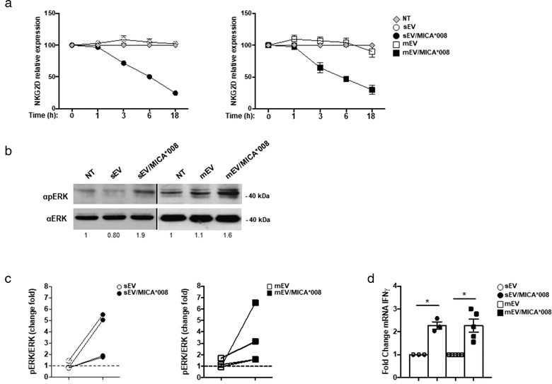
FIGURE 6.

MICA*008 positive EVs engage NKG2D and trigger pERK and IFN‐γ production. (a) NKL cells were treated for different times with MICA*008+ or MICA*008– sEVs (at 30 μg/ml) or mEVs (at 20 μg/ml). Cells were harvested and NKG2D expression was evaluated by immunofluorescence and FACS analysis. Relative expression of NKG2D was calculated considering the untreated group as 100%. (b) NKL cells were treated for 1 h with MICA*008+ or MICA*008– EVs. Western blot analysis was performed on total cell lysates using ERK and phospho‐ERK (pERK) Abs. Numbers beneath each lane represent quantification of pERK by densitometry analysis normalized with ERK relative to the untreated group. (c) Quantification of immunoblots relative to different independent experiments is shown. Dashed line represents the untreated group. (d) Highly purified human primary NK cells were incubated with 30 μg/ml of sEVs or 20 μg/ml of mEVs expressing or not MICA*008 for 24 h. Real‐time PCR analysis of IFN‐γ mRNA is shown. Data, expressed as fold change units, were normalized with β‐actin and referred to MICA*008– EVs treated cells considered as calibrator. Values reported represent the mean of at least three independent experiments
