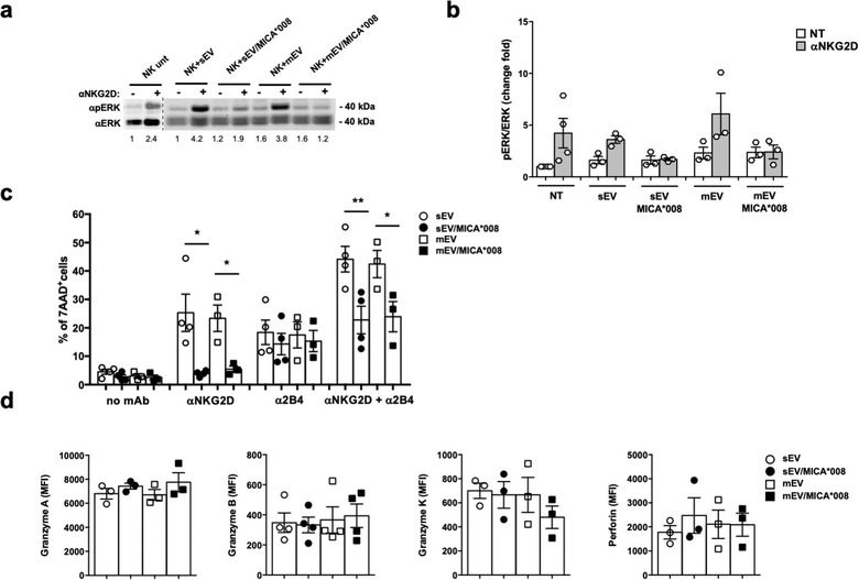
FIGURE 7.

Prolonged exposure to MICA*008+ EVs impairs both NKG2D signalling pathway and NKG2D‐mediated killing. NKL cells were treated for 24 h with 30 μg/ml of sEVs or 20 μg/ml of mEVs expressing or not MICA*008. (a) Untreated or EV‐treated NKL cells were stimulated with anti‐NKG2D mAb plus goat anti‐mouse IgG for 15 min at 37°C before the lysis. Western blot analysis was performed on total cell lysates using ERK and phospho‐ERK (pERK) Abs. Numbers beneath each lane represent quantification of pERK by densitometry analysis normalized with ERK relative to the untreated group. (b) Quantification of immunoblots relative to independent experiments is shown. (c) The cytotoxic activity of EV‐treated NKL cells was evaluated by performing the 7‐AAD assay in a redirected modality against P815 FcR+ target cells loaded with different Ab as indicated in figure, at an E:T ratio of 25:1. Data reported represent the mean of at least three independent experiments. d) The NKL intracellular amount of perforin, granzyme A, B and K was evaluated through immunofluorescence and FACS analysis. The mean of at least three independent experiments is shown. Values represent the MFI of granzyme A, B, K and perforin subtracted from the MFI value of the cIg
