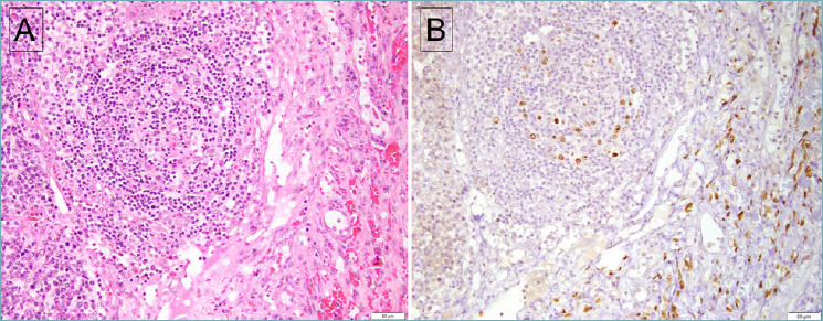
Figure 1.

Lymph node involved by both MCD and KS. (A) The follicle shows regressive changes, with an adjacent spindle cell proliferation associated with extravasated red blood cells. (B) Staining for LANA-1 is positive in both lymphoid cells within the follicle as well as the spindle cells of KS. However, the spindle cells of KS are negative for vIL-6 (not shown).
