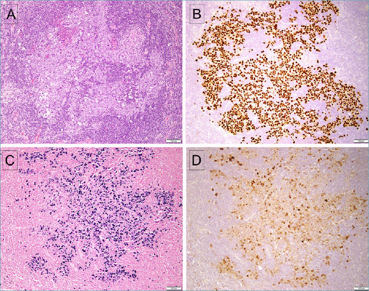
Figure 3.

Germinotropic lymphoproliferative disorder. (A) The affected follicle shows irregular cords and sheets of atypical large lymphoid cells. (B) The lesional cells are positive for LANA-1, and also positive for EBV, with EBER in situ hybridization (C). D. The affected cells are also positive for vIL-6.
