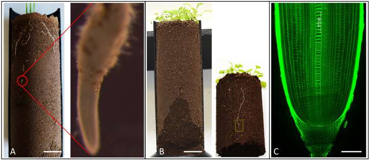
Figure 3. Identification and selection of roots for anatomical scale imaging.
Rice (A) and Arabidopsis root (B) grown in 3D printed (half open) column growing in compacted soil. The red circle and yellow rectangle show the root tip zone of rice and Arabidopsis roots, respectively. (C) Longitudinal image of rice root tip grown in non-compacted soil.

