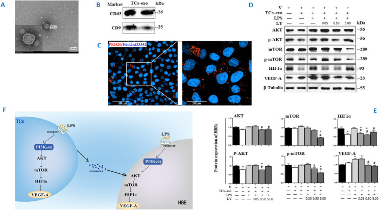
Fig. 6.
Roles of TC-derived exosomes in HBE response. The ultrastructure of exosomes was identified (A) using TEM (40, 000 ×) and CD81, CD61, and CD9 of exosomes were measured by Western blot (B). The distributions of PKH26-labeled exosomes were monitored with a confocal microscope (63 × water mirror) (C). Expression of AKT, p-AKT, mTOR, p-mTOR, HIF1α, VEGF-A, and MMP9 protein was assayed (D and E). The bands in D are WB bands. The result of quantitative analysis of WB bands is shown in E. Potential mechanisms were proposed to be responsible for the intercellular communication (F). * and ** stand for p values less than 0.05 and 0.01, as compared with HBE pretreated and challenged with vehicle (V), while # and # # stand for p values less than 0.05 and 0.01, as compared with cells pretreated with vehicle and challenged with LPS + TCs-exo, respectively

