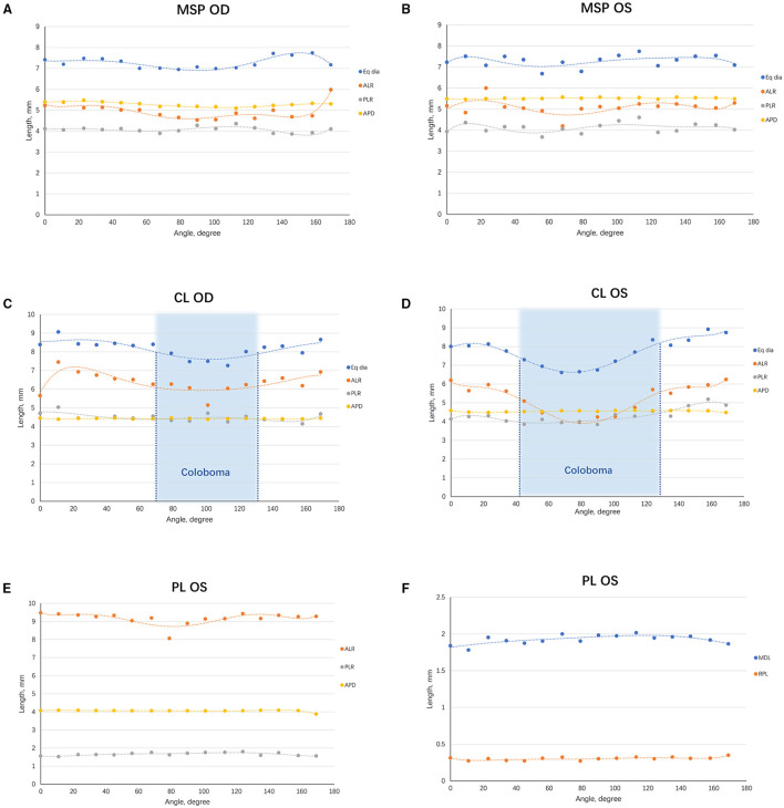
Figure 4.
Scatterplots demonstrating the trend of lens biometric parameters from various angles. (A) The lens biometric parameters of the right eye of MSP at different angles were plotted and fitted in curves. (B) A similar graph was made on the left one. (C) The lens biometric parameters of the right eye of CL at different angles were plotted and fitted in curves. The coloboma ranged from 250° to 311° (indicated in the blue area). (D) A similar graph was made on the left one. A larger range of coloboma was observed in the left eye (222° to 308°, indicated in the blue area). (E) The ALR, PLR, and APD of the eye with PL. (F) The MDL and RPL of the eye with PL. ACD, anterior chamber depth; ALR, anterior lens radius; APD, anteroposterior distance; CL, coloboma lentis; Eq Dia, equatorial diameter; MDL, maximum diameter of the lesion; MSP, microspherophakia; PL, posterior lenticonus; RPL, rear projection length; PLR, posterior lens radius.

