Fig. 6.
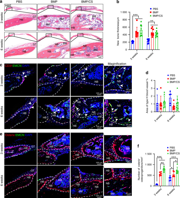
Induced PTs enhance osteogenesis and osseointegration during autologous transplantation for calvarial defect repair. a, b H&E staining (a) and quantitative analysis (b) were used to assess the integration of transplanted PTs with host bones (HBs) (n = 8). For PT in the PBS group, only new bone differentiated from HBs was observed. For PT in the BMP group, the mineralized HBs and the newly generated bones were partly separated by soft fibrous tissue. In contrast, HBs completely integrated with newly generated bones differentiated from the transplanted PTs in the BMP/CS group. c–f Immunofluorescence staining and quantitative analysis of type H blood vessels (EMCN + CD31 +, yellow; c, d) and Osterix+ osteoprogenitors (red; e, f) in the PBS, BMP, and BMP/CS groups at weeks 3 and 6 after autogenic transplantation in young mice. Bone marrow BM, new bone NB, host bone HB, granulation tissue GT. EMCN-positive cells are green, CD31- and Osterix-positive cells are red, and DAPI is blue (nucleus). The white arrowhead indicates type H blood vessels (c). The white dashed line indicates the host bone or new bone area (c, e). Scale bar (top: low magnification, 1 mm; bottom: high magnification, 200 μm; a) and (left: low magnification, 100 μm; right: high magnification, 50 μm; c, e). Data are presented as the mean ± SD. *P < 0.05, **P < 0.01, ***P < 0.001, two-way ANOVA followed by Bonferroni’s post hoc tests

