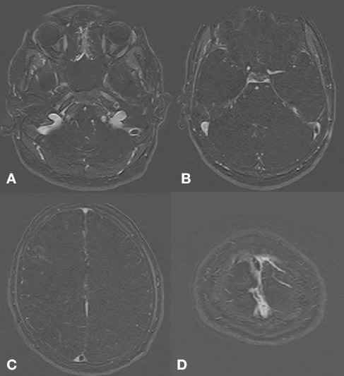
Figure 2.

Filling defect is identified in the left sigmoid sinus (A) extending into the distal transverse sinus (B). There is also a filling defect in the torcula Herophili (C) extending into the distal portion of the superior sagittal sinus. Partial filling defect is also identified in the superior sagittal sinus near the vertex (D). Findings are suggestive of cerebral venous sinus thrombosis.
