FIGURE 3.
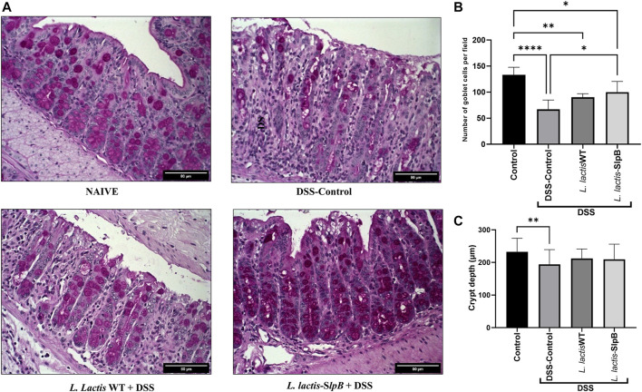
L. lactis-SlpB mitigates histological signs of DSS-colitis. Model images of micrographs for analysis of goblet cells in colon tissue (A) and the result of goblet cell quantification by field (B) and depth of colon intestinal crypts (C) are shown. The slides were stained in Periodic Acid-Schiff (PAS), goblet cells have intense purple-pink tones, and analyzed under ×40 magnification. The data represent the mean ± SD of 12 mice per group. One-way ANOVA and Tukey’s post-hoc tests were used for multiple comparisons. Asterisks represent statistically significant differences as follows: ∗p < 0.05, ∗∗p < 0.01, ∗∗∗p < 0.001, ∗∗∗∗p < 0.0001.

