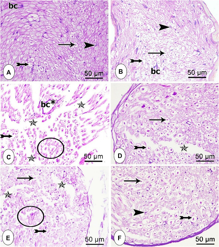
FIGURE 3.
Microscopic representative images of the sciatic nerve transverse sections from the different studied groups (A) control group; (B) sham group (C) CCI group; (D) CCI + pregabalin, (E) CCI + P. perfoliatus extract (200 mg/kg, p.o) (F) CCI + P. perfoliatus extract (400 mg/kg, p.o). Arrow, bifid arrow, arrowhead, star, and circle illustrate axoplasm, area of dissolved myelin, Schwann cells nuclei, wide separation between the nerve fibers, and degenerated nerve fibers, respectively. (bc) blood vessel, and (bc*) dilated blood vessel. Scale bar, 50 μm x400.

