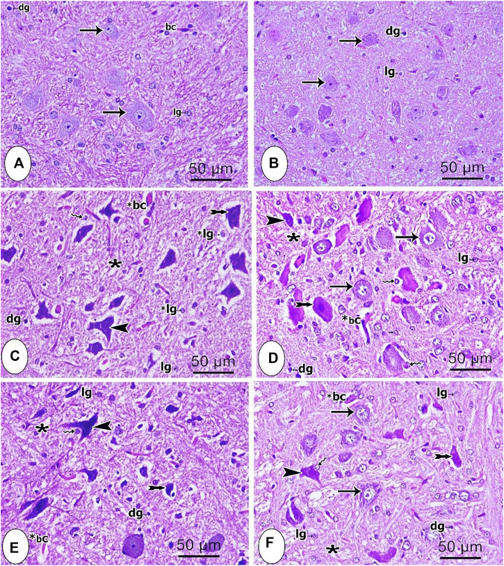
FIGURE 4.
Microscopic images of brainstem tissues from the different studied groups (A) control group (B) sham group; (C) CCI group (D) pregabalin group; (E) P. perfoliatus extract (200 mg/kg, p.o.) (F) P. perfoliatus extract (400 mg/kg, p.o.). Arrow, arrowhead, bifid arrow, wavy arrow, and (*) represent normal neurons with flame elongated dark nuclei, neurons with dark stained nuclei and vacuolated cytoplasm, perineural glial cell nuclei, and neuropil respectively. (bc) normal blood capillary, (*bc) dilated blood capillary, (lg) lightly stained glial cell nuclei, (dg) deeply stained glial cell nuclei. Scale bar, 50 μm x400.

