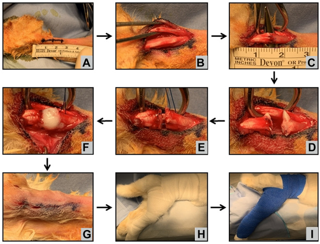
Figure 1.
Surgical technique demonstrating the rabbit positioned prone with the hindlimb prepared and draped. (A) A 2-cm incision centered 1.5 cm proximal to the calcaneal tubercle was performed. The paratenon was incised, and the (B) flexor digitorum superficialis and (C) Achilles tendon were identified and isolated. (D) Tenotomy was made through the Achilles tendon. (E) A modified Kessler core suture was performed in all groups. (F) The scaffold was placed at the tenotomy site for groups 2 and 3 before final suture tightening. (G) The incision was closed using absorbable suture, and (H, I) the hindlimb was placed in a hip spica–like cast at 150° for 3 to 6 weeks.

