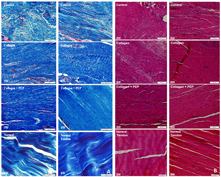
Figure 4.
(A) Trichrome and (B) hematoxylin and eosin stains of specimens from each group at each time point as well as normal contralateral, untreated tendon. Images show more organized, denser collagen with less peripheral adhesions in the purified exosome product (PEP)–treated group, more closely resembling normal tendon.

