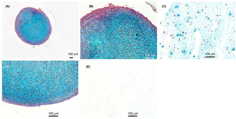
Figure 6.
Histology sections of chondrocytes differentiated for 2 weeks. (A-C) 3D bioprinted primary chondrocytes in 80:20 NFC:A bioink followed by differentiation for 2 weeks. Micro tissue structures similar to pellets can also be seen in the 3D prints. (D) Control primary chondrocytes differentiated as micromass pellets for 2 weeks. (E) Control print 80:20 NFC:A bioink with no cells. Scale bars, 100 µm.

