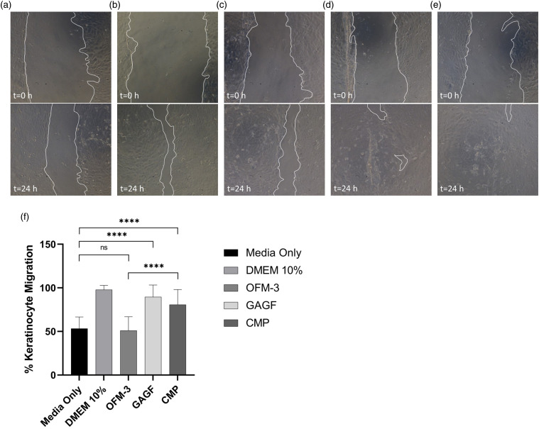
Figure 7.
In vitro human keratinocyte migration. Representative images of scratched mono-layers at t = 0 h and t = 24 h for the test samples, media only (a), DMEM +10% serum (b), OFM-3 (c), HA (d), and CMP (e), (f) Quantification of average percent epithelial migration. Error bars represent standard deviation from triplicate independent experiments with n = 6 sample replicates per experiment. ****, p < .0001; ns = not significant. DMEM: Dulbeccós modified Eaglés Medium; OFM: Ovine forestomach matrix-3; CMP: composite samples.

