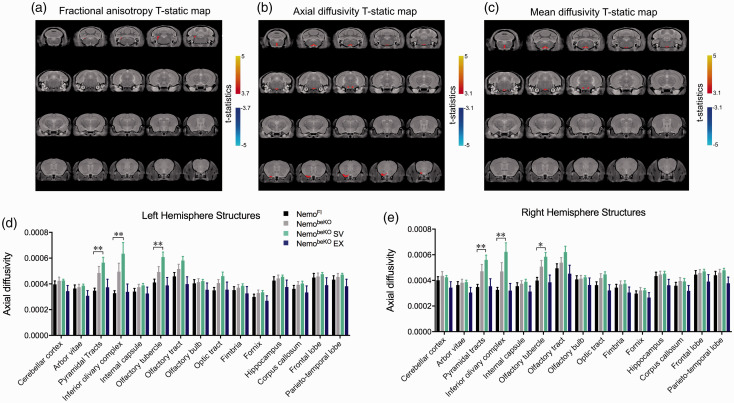
Figure 4.
Diffusion tensor imaging analysis from magnetic resonance imagining images including fractional anisotropy (A), axial diffusivity (B), and mean diffusivity (C) with statistical threshold shown whereby NemobeKO mice treated with simvastatin (SV) are significantly different in both left (D) and right hemisphere (E) structures including the pyramidal tract, olfactory tubercle, and inferior olivary complex. One-way ANOVA with post-hoc Newman-Keuls multiple comparison tests were performed for each structure of interest for axial diffusivity. N = 8–10 mice/group, *p < 0.05 and **p < 0.01.

