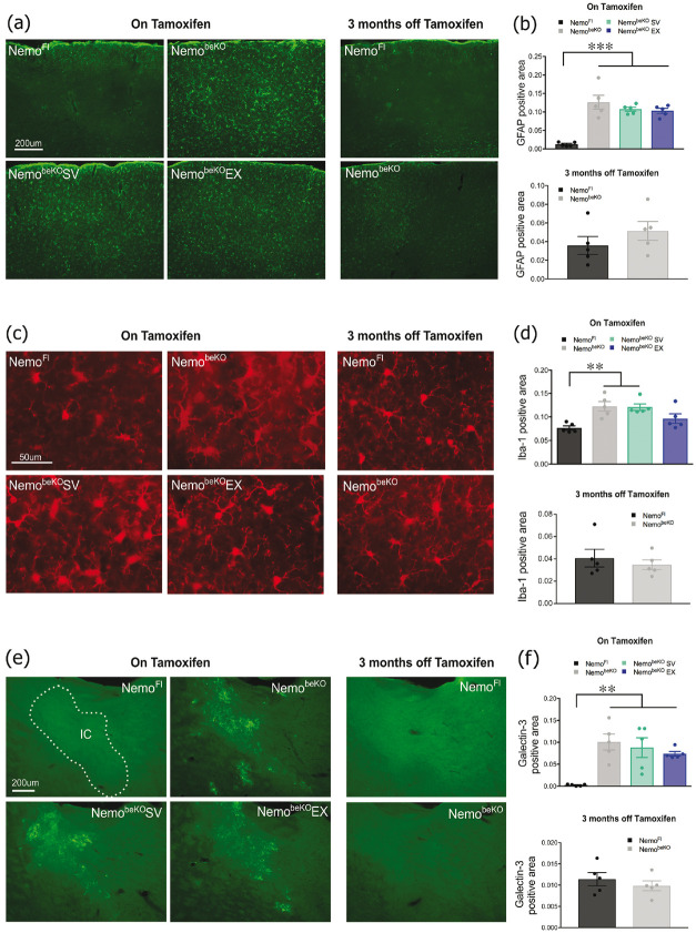
Figure 5.
Astrogliosis detected with GFAP immunofluorescence was significantly increased in the cortex of all NemobeKO groups regardless of treatment but returned to normal in the recovery cohort (A and B). Microglial activation in the cortex was also significantly raised in the NemobeKO groups as quantified by Iba-1 positive area, with the exception of mice that exercised (EX) and returned to normal in the recovery cohort (C and D). Inflammation in the internal capsule (IC) was increased in all NemobeKO groups identified by galectin-3-positive microglial cells, a response completely normalized in the recovery cohort (E and F). N = 4–5 mice/group. One-way ANOVA with post-hoc Newman-Keuls multiple comparison tests were performed **p < 0.01, ***p <.001.

