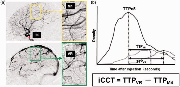
Figure 1.
Measurement of iCCT.
A, ROIs were set in the vertical intracavernous portion of the internal carotid artery (C5), the cortical segment of the rolandic artery (M4), and the rolandic vein (VR) on the images of lateral projection. B, Time-density curve of the contrast media in each ROI representing the various times to peak (TTP), and intracerebral circulation time (iCCT).

