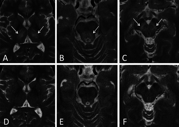
FIG 1.

Axial T2‐weighted brain MR images showing pre‐treatment abnormal bilateral and symmetrical hyperintensities in the lateral thalamus (A), central midbrain (B) and substantia nigra (C) (white arrows) with post‐treatment complete resolution of abnormalities (D, E, F). The hypointensities discussed in the text are not visible in these images.
