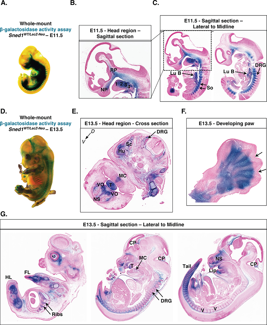
Figure 6. Patterns of expression of Sned1 gene during embryogenesis.
A. Whole-mount β-galactosidase assay (LacZ staining) performed on heterozygous E11.5 Sned1WT/LacZ-Neo embryo.
B. Sagittal section of the head region of LacZ-stained E11.5 LacZ Sned1WT/LacZ-Neo embryo. 1, 2, 3, and 4 represent the branchial arches; NP: nasal process; RP: Rathke’s pouch.
C. Sagittal sections of LacZ-stained E11.5 LacZ Sned1WT/LacZ-Neo embryo (left panel: lateral section; right panel: across the midline). Lu B, lung bud; So, somites; DRG, dorsal root ganglia.
D. Whole-mount β-galactosidase assay (LacZ staining) performed on heterozygous E13.5 Sned1WT/LacZ-Neo embryo.
E. Transverse section of the head region of LacZ-stained E13.5 LacZ Sned1WT/LacZ-Neo embryo. DRG, dorsal root ganglia; Hu: humerus; MC: site of apposition in the midline of Meckel’s cartilages; NS, cartilage primordium of nasal septum; Sc: blade of scapula; T, tongue; VO, vomeronasal organ.
F. Section of paw from LacZ-stained E13.5 LacZ Sned1WT/LacZ-Neo embryo. Arrows indicate interdigital spaces.
G. Sagittal sections of Lac-Z-stained E13.5 LacZ Sned1WT/LacZ-Neo embryo (from left to right: lateral to the midline). CP, choroid plexus; DRG, dorsal root ganglia; FL, forelimb; HL, hindlimb; NS, cartilage primordium of nasal septum; T, tongue; V, cartilage primordium of the vertebrae.

