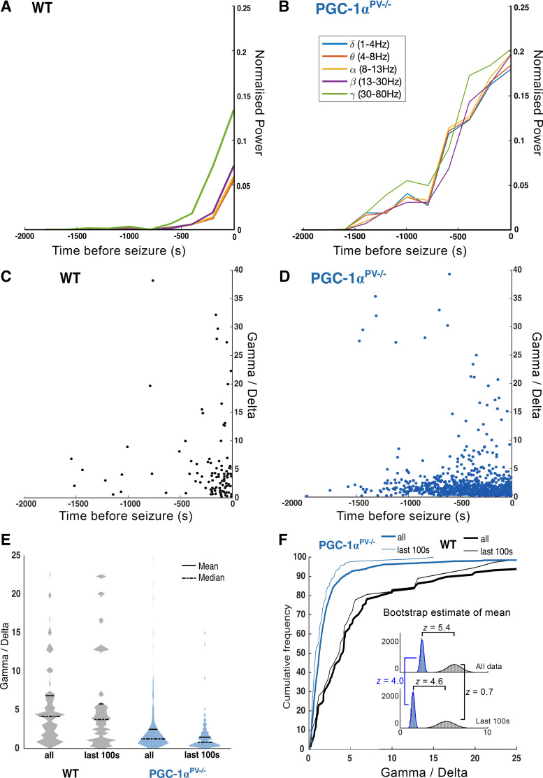
Figure 4.
Preictal discharges in brain slices from PGC-1α knockout mice show reduced normalized gamma activity (γ/δ ratio) compared with wild-type (WT) mice. A: the mean normalized power, for the main physiological LFP bandwidths as indicated, averaged for sequential 200 s epochs before the onset of the first SLE (t = 0 s), for wild-type brain slices, recorded in 0 Mg2+ ACSF (n = 7). B: equivalent data from brain slices from PGC-1αPV−/− mice (n = 10). Note the far more rapid escalation of activity in the wild-type brain slices, especially in the gamma and high-gamma frequency bands, leading up to the first SLE. The γ/δ ratio for every single interictal event, plotted against time relative to the onset of the first SLE, in the wild-type brain slices (C), and in the PGC-1αPV−/− brain slices (D). E: violin plots of γ/δ ratios, for the entire data set of interictal events, and, events occurring in the 100 s immediately before the first SLE. F: cumulative frequency plots of the same data sets. Insets: the probability distribution for estimates of the means for the full data sets, and the events within 100 s before the first SLE, derived by bootstrap resampling with replacement. Z-scores are provided for the four group comparisons, all except the wild-type, all-vs.-last 100 s comparisons are highly significant.

