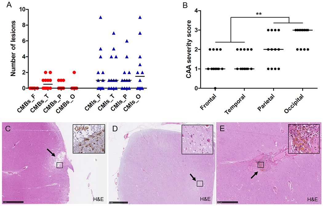
Figure 3.

Regional associations on standard histopathologic examination of microbleeds and microinfarcts with CAA severity.
On average, CAA severity across all 12 cases followed an anterior-to-posterior distribution (B), whereas number of histopathologically-observed microbleeds and microinfarcts did not (A), explaining weak regional associations. From the total number of 94 microinfarcts observed on standard histopathology, 61 (65%) were considered old/chronic based on GFAP positivity (inset) (C, this example follows the perfusion area of a penetrating cortical vessel) and 33 (35%) recent/acute based on the presence of ‘red’ neurons (inset) (D, this example is located deeper (within cortical layer III-VI)). From the total number of 13 microbleeds observed on standard histopathology, 12 (92%) were considered old/chronic based on the presence of hemosiderin-containing macrophages (inset) (E), and 1 (8%) recent/acute (not shown). Median is indicated in A and B. Scale bar in C and D = 1 mm, scale bar in E = 500 μm.
