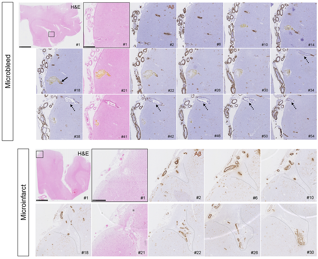
Figure 7.

Serial H&E and Aβ stained sections capturing a recent/acute microbleed and an old/chronic microinfarct.
Vessels responsible for microbleeds were traced on serial sections, stained with H&E (#1, 21, 41 etc.) and Aβ (#2, 6, 10 etc.) and revealed absence of Aβ at the rupture site, but subtle Aβ upstream and downstream (arrow). This example is a recent/acute microbleed characterized by hematoidin (yellow substance). Note that the vessel is enlarged at the site of bleeding (section #21). Moreover, this vessel did not have any SMCs left but showed extensive fibrin(ogen) build-up in the wall (not shown). Note the vessel that runs in parallel to the microbleed (broken arrows), which is also abnormally enlarged and shows loss of Aβ from the vessel wall but has not ruptured. Scale bar in first panel = 5 mm, scale bar in second panel = 500 μm. Microinfarcts were traced on serial sections, stained with H&E (#1, 21, 41 etc.) and Aβ (#2, 6, 10 etc.), which revealed extensive vascular Aβ at the core of the microinfarcts, as well as upstream and downstream. This example is a chronic/old microinfarct characterized by tissue loss, cavitation, and GFAP positivity (not shown). Note that the walls of the vessels at the core of the microinfarct appear relatively intact, except that they lost their SMCs (not shown). Scale bar in first panel = 5 mm, scale bar in second panel = 500 μm.
