Fig. 5.
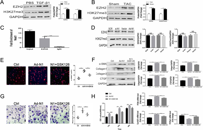
Aggravation of cardiac fibrosis by Neat1 via recruitment of EZH2. A, B Representative western blotting and quantitative analysis findings for EZH2 and H3K27me3 in CFs (n = 5 in each group). C Interaction between Neat1 and EZH2 verified by the RIP assay (n = 5 in each group). D Representative western blotting and quantitative analysis findings for EZH2 and H3K27me3 in CFs (n = 5 in each group). E CFs were analyzed by immunofluorescence for the expression of α-SMA (red) and nuclei (DAPI: blue) (n = 5 in each group; scale bar = 500 µm). F Representative western blotting and quantitative analysis of α-SMA and ECM-related proteins in CFs (n = 5 in each group). G Representative images of Transwell migration assay and quantification of migrated CFs (n = 5 in each group; scale bar = 100 µm). H Quantification of cell proliferation by the CCK-8 assay (n = 5 in each group) and mRNA levels of PCNA, cyclin D1, and P27 Kip1 by qRT-PCR. Data are presented as mean ± SEM. *p < 0.05, **p < 0.01, ***p < 0.001, NS = no significant difference between the indicated groups

