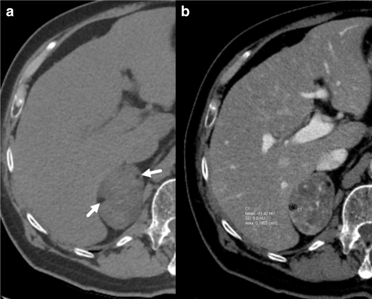
Figure 1.
62-year-old female with a clinical diagnosis of Cushing’s syndrome. Axial unenhanced CT (a) demonstrates a large right adrenal nodule containing small foci of fat attenuation (arrows). Postcontrast imaging (b) demonstrates heterogeneous nodule enhancement with a Hounsfield unit (HU) of −51 in one of the foci confirming macroscopic fat. The nodule was later pathologically confirmed to be an adrenal adenoma with foci of myelolipomatous degeneration.

