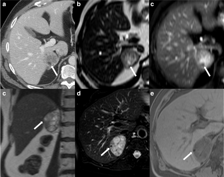
Figure 5.
52-year-old female with an incidentally discovered right adrenal nodule and subclinical Cushing syndrome. Contrast-enhanced axial CT through the upper abdomen (a) demonstrates a right adrenal nodule containing a tiny focus of macroscopic fat (arrow). Axial MRI T2-weighted (b) and T2-weighted fat-saturated (c) images show the same focus (arrow) lose signal with fat saturation, confirming the presence of macroscopic fat. An additional focus of fat seen on the coronal T2-weighted image (d) also shows loss of signal on fat-saturated T2-weighted (e) and fat-saturated T1-weighted (f) sequences. The nodule was pathologically proven as an adrenal adenoma with foci of myelolipomatous degeneration.

