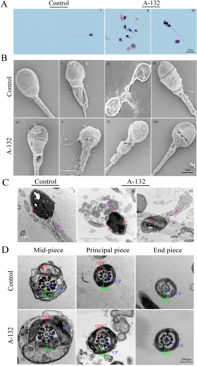
Fig. 2.

Sperm morphology and ultrastructure analyses in the spermatozoa from SLO3-mutated subject A-132. A Light microscopy analysis of spermatozoa from control (i) and the individual harbouring the SLO3 variant. Most spermatozoa from the individual harbouring the SLO3 variant have small acrosomal heads, swollen midpieces, and coil-shaped flagella. B SEM analyses of sperm cells from a fertile control and the SLO3-mutated individual (II-VIII). Spermatozoa of normal controls exhibit a smooth, regularly contoured, oval-shaped head and a flagellum with a clearly defined midpiece, whereas most sperm from the SLO3 mutant individual exhibit severe morphological defects (II-VIII), such as swollen midpieces (II), coil-shaped flagella (III), small acrosome (IV), and absence of acrosome in combination with a defective midpiece (V-VIII). C TEM analyses of sperm cells from a fertile control and the SLO3-mutated individual. The longitudinal sections of spermatozoa from control and A-132. Sperm tails with poorly assembled mitochondria (II-red arrow) or a cytoplasmic mass containing different components of the flagellum (III-red arrow) are observed. The acrosome is thin or broken with unidentifiable acrosomal membranes (red asterisk) along with misshapen heads; chromatin condensation appears abnormal (number sign). D Cross-sections of the mid-piece, principal piece, and end piece of the flagella in a control individual show the typical “9 + 2” microtubule structure, including 9 peripheral microtubule doublets paired with 9 outer dense fibres and the central pair of microtubules, surrounded by the organized mitochondrial sheath or fibrous sheath. Ultrastructures of sperm from the SLO3-mutated individual are comparable to those from control. Scale bars: 10 μm (A), 1 μm (B), 2 μm (C), and 200 μm (D)
