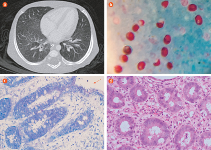
Figure 1.
(a) CT scan of the chest showing scattered faint ground-glass appearance related to non-specific pulmonary infection. (b) Cryptosporidium oocyte in acid-fast stain. Oocysts appear as red, oval-shaped, in blue/green background 100 ×. (c) A modified Ziehl–Neelsen stain on low power showing the Cryptosporidium oocytes within the crypts and over the epithelial surface (red arrow) 20 ×. (d) Hematoxylin and eosin stain of colonic tissue on high power reveal features of colitis and the presence basophilic spherical organisms within the crypts, morphologically consistent with Cryptosporidium 20 ×.

