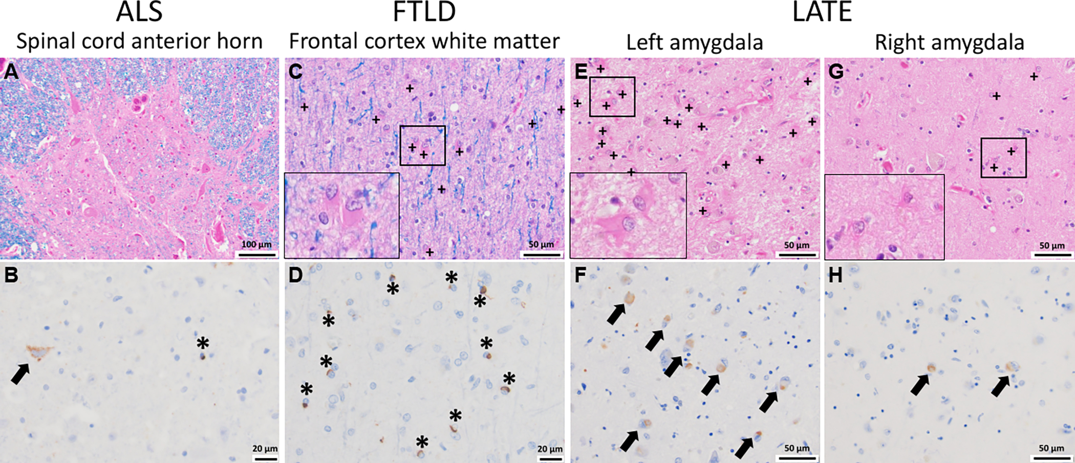
Figure 2. TDP-43 pathology and glial responses across the TDP-43 proteinopathies.

(A, B) In ALS, cells within the anterior horn of the spinal cord are affected (A, H&E/LFB stain) and immunohistochemical staining for phosphorylated TDP-43 highlights inclusions both in neurons (arrow) and glia (asterisk) (B, pTDP-43 immunostain). (C, D) In FTLD, the subcortical white matter is often gliotic as evidenced by increased reactive astroglia (plus signs and inset) (C, H&E/LFB stain) in association with numerous pTD-43 inclusions in oligodendroglia (asterisk, D, pTDP-43 immunostain). (E-H) In LATE, which is often seen comorbid with Alzheimer’s disease, there is a robust glial reaction (E, G, H&E/LFB stain, with reactive astrocytes shown by plus signs and in the insets) in association with the presence of neuronal pTDP-43 inclusions (arrows, F, H, pTDP-43 immunostain). In this case, while pTDP-43 inclusions were found bilaterally in the amygdala, the left side showed significantly more inclusions compared to the right, which correlated with the striking increase in reactive astroglia also seen in the left amygdala (inset). Whether the pTDP-43 inclusions are found in glia as well as neurons (arrows) is not well characterized in the context of AD+LATE. + = reactive astrocyte, arrow = neuronal TDP-43 inclusion, * = glial TDP-43 inclusion
