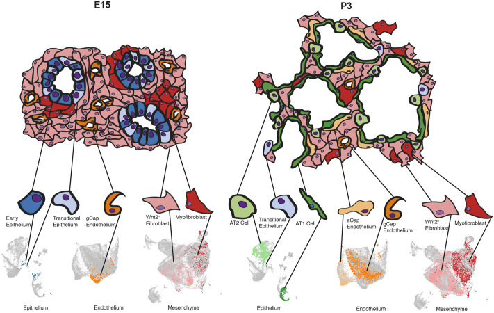
Fig. 7.
Functional development of the lung is a result of spatial and temporal organization of multiple cell types. An abbreviated summary model of the developing lung compared between two representative time points: mid-embryogenesis (E15) and postnatal (P3). At E15, the lung epithelium is composed of indistinct, uncommitted cell types (as defined by marker-gene analysis) that are very rare or absent in adulthood. The prenatal endothelium in the lung parenchyma is composed of general capillary (gCap) endothelial cells, whereas the majority of cells in the remaining tissue are Wnt2+ fibroblasts, with interspersed clusters of myofibroblasts. By P3, many cell types observed in mature lungs are already present, including AT1, AT2 and transitional epithelial cells. The gCap endothelium has differentiated into Car4+ alveolar capillary (aCap) cells, alongside the gCap endothelium, and mesenchymal cells have undergone extensive spatial reorganization. Representative cell clusters are highlighted in the inset UMAPs derived from Fig. 1C.

