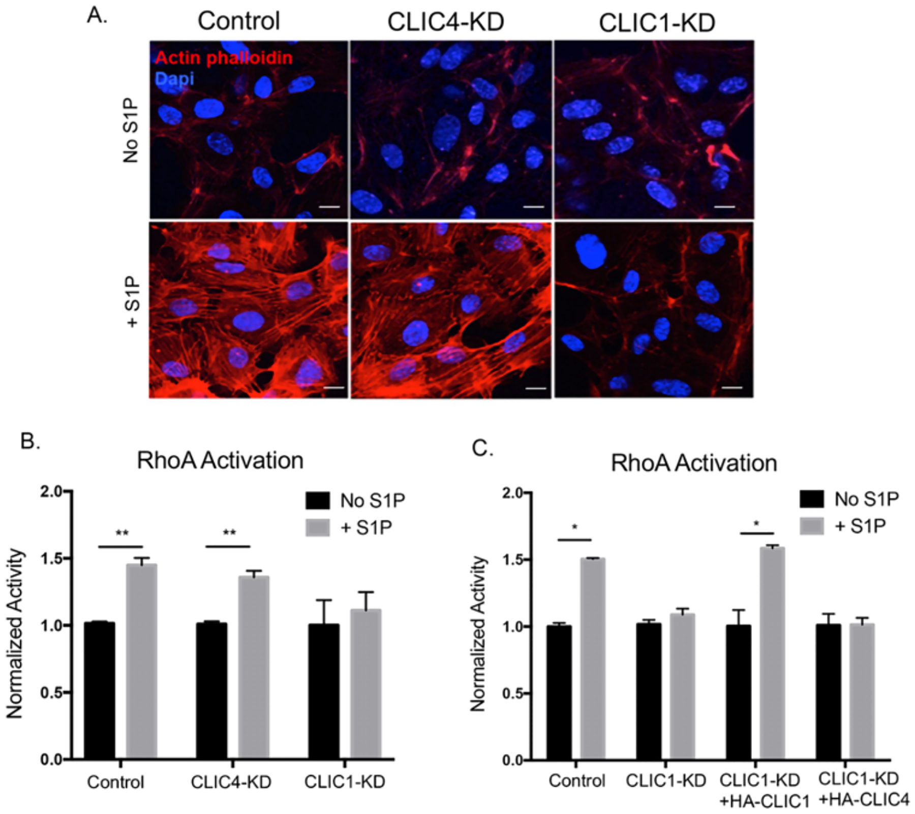
Figure 5. CLIC1 regulates endothelial S1P-driven RhoA activity.

(A) HUVECs were serum starved for 3hrs then stimulated with 1μM S1P for 30min. Cells were fixed and stained with phalloidin to visualize actin stress fibers (red). DAPI was used as a nuclear stain (blue). Confocal images are representative of three independent experiments. Scale bar = 10 μm (B and C) HUVECs were serum starved for 3hrs then treated with 1μM S1P for 5min. RhoA activity was measured by G-LISA assay and normalized to unstimulated cells for each condition (ANOVA with Bonferroni correction for multiple comparisons test, N=3, *p<0.05, **p<0.01).
