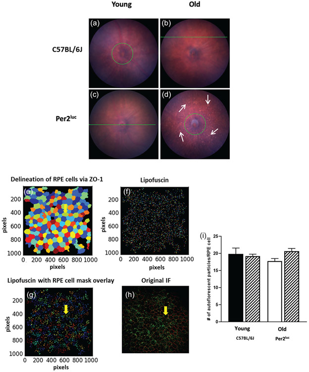
Figure 3. Aged Per2luc mice display some abnormality in the fundus, but no difference in RPE cell apical autofluorescence.
At the younger age, no differences were observed in the fundus image between the two genotypes (A, B) whereas at older age we observed an increase in the number of fundus autofluorescence spots (white arrows) in Per2luc (D) when compared to C57BL/6J of similar age (C). We then counted the number of apical autofluorescent particles present in isolated RPE cells in RPE flat mount preparation using cell profiler (v 2.2.0) which allowed the delineation of RPE cells via ZO-1 staining (E) and autofluorescent particles (i.e., lipofuscin) (F). Finally an RPE cells ZO-1 mask was created which was then overlaid on the isolated autofluorescent particles (G). When the lipofuscin particles were counted in each isolated RPE cell, there were no differences observed in the number of autofluorescence particles (I; p > 0.05, two-way ANOVA, Tukey post-hoc, n=3-4). A representative immunofluorescence image of a typical RPE flat mount with ZO-1 and autofluorescent particles from a young C57BL/6J mouse (H).

