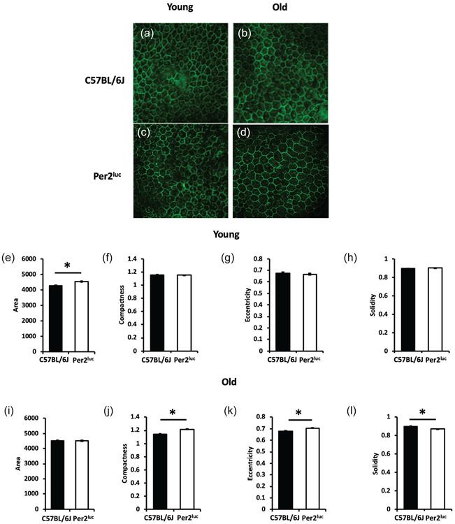
Figure 4. Aged Per2luc mice show signs of premature aging in the RPE.
RPE cell shape morphology was assessed in young C57BL/6J (A) and Per2luc (C) mice and old C57BL/6J (B) and Per2luc (D). We observed an increase in total RPE cell area in young Per2luc mice when compared to similarly aged C57BL/6J mice (E; *=p < 0.05, two-way ANOVA, Tukey post-hoc, n=3-4) with no change in the other cell morphology parameters measured at this age (F, G, H; p > 0.05, two-way ANOVA, n=3-4). However, when these mice were old, there was no longer any difference in total RPE cell area (I; p > 0.05, two-way ANOVA, n=3-4) but there was an increase in compactness, eccentricity, and a decrease in solidity when compared to age-matched C57BL/6J mice (J, K, L; *=p < 0.05, two-way ANOVA, Tukey post-hoc, n=3-4). Photomicrographs show representative RPE flat mount images of ZO-1 cell junction protein demarcating RPE cell boundaries in C57BL/6J and Per2luc mice at both young and old age.

