Figure 1.

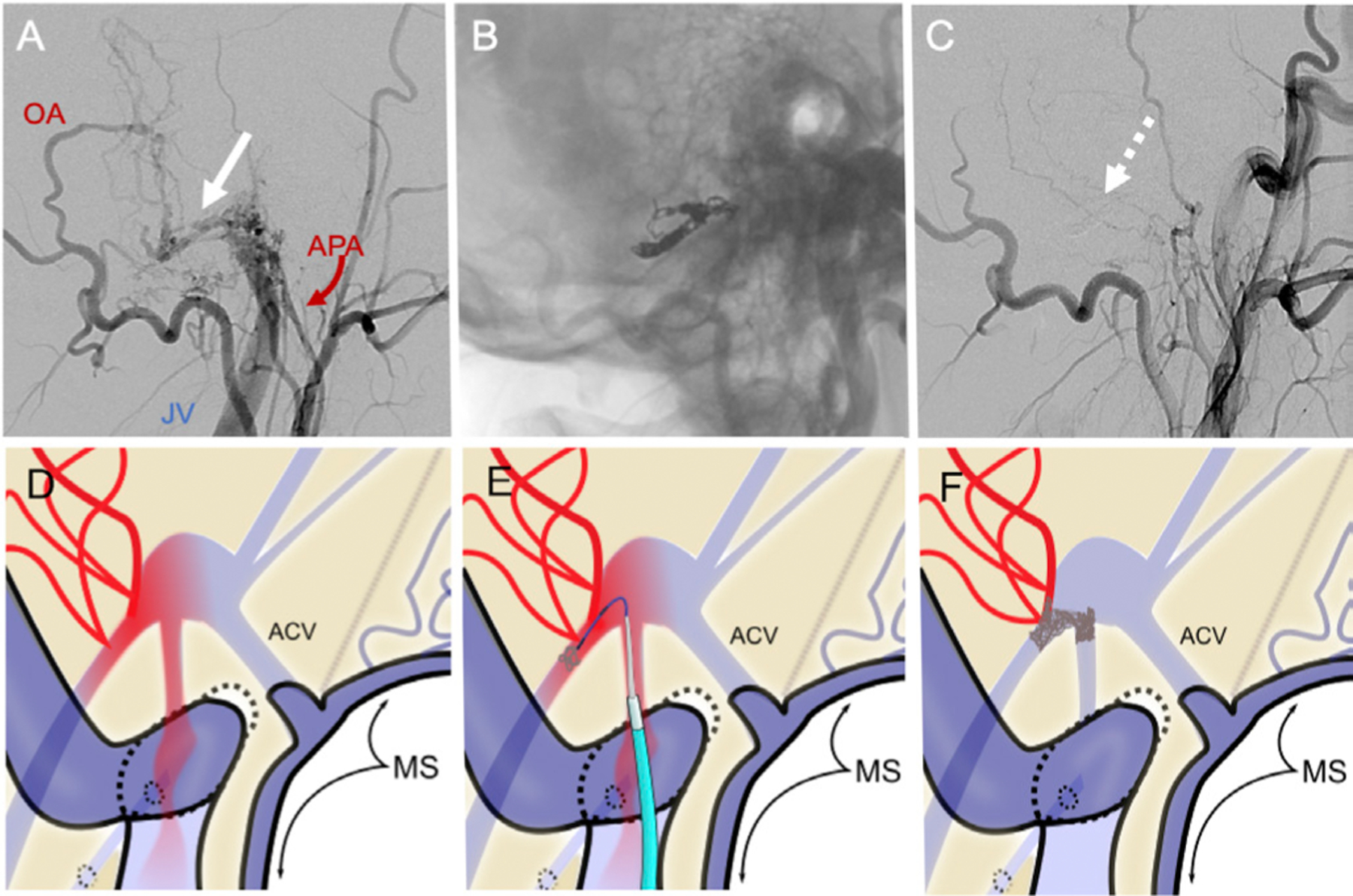
Pretreatment left external carotid artery injection (A, lateral projection) shows lateral condylar vein dural arteriovenous fistula (white arrow) supplied by the occipital artery (OA) and branches of the ascending pharyngeal artery (APA) with early filling of the jugular vein (JV). The fistula was treated with transvenous embolization of the venous pouch (B). Post-treatment angiogram (C) shows complete resolution of the fistula (dashed arrow). (D–F) Schematic depiction of triaxial transvenous (TV) approach to the foramen magnum region-dural arteriovenous fistula (FMR-AVF): Part D shows salient anatomy of an FMR-AVF (arterial blood=red), including the marginal sinus (MS) and anterior condylar vein (ACV); the direction of antegrade flow in low-risk AVF. Catheter positioning for TV coil embolization is shown in (E), and postembolization angiographic cure is depicted in (F).
