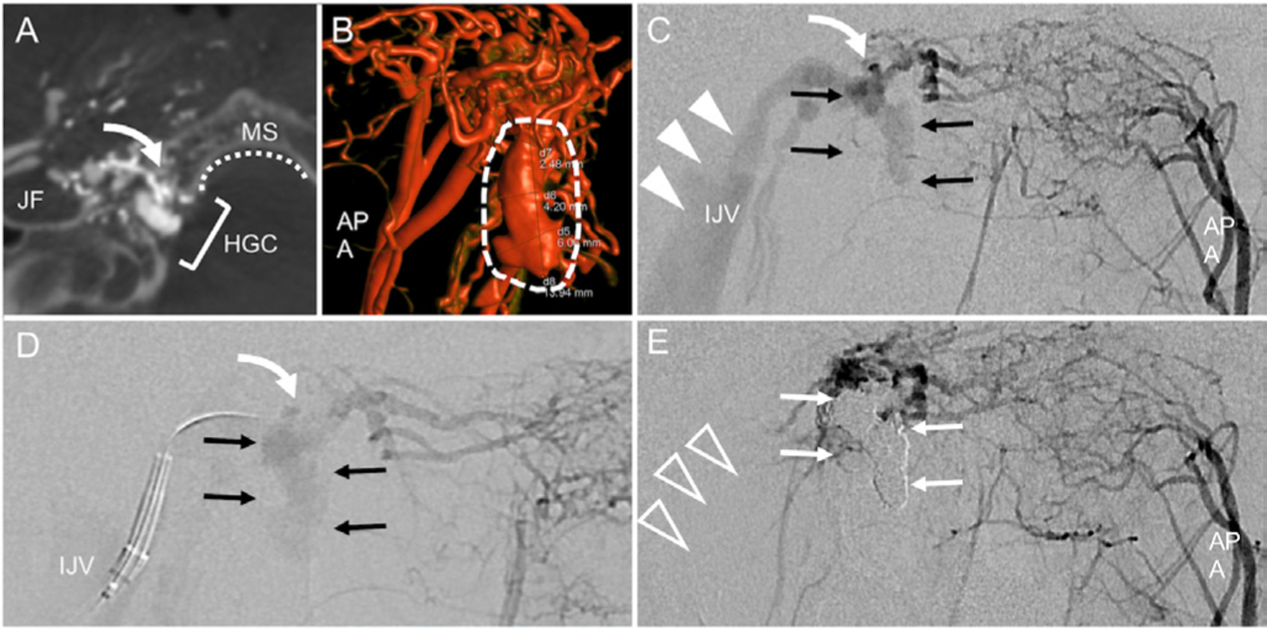
Figure 2.

Anterior condylar vein arteriovenous fistula. Dyna-CT angiography with injection of the right ascending pharyngeal artery (APA) shows anatomy of the shunt (curved arrow) within the hypoglossal canal (HGC), adjacent to the marginal sinus (MS) and jugular foramen (JF). 3D re-formation shows that the fistulous pouch (dashed arrows) extends caudally into the MS. Panels C-E show sequential steps of arteriography-guided transvenous embolization: Water’s/anteroposterior (AP) view of left APA injection shows the fistula (white curved arrow) and venous pouch (black arrows) with the ipsilateral jugular vein (IJV, white arrowheads). In D, a guide catheter is positioned in the IJV with microcatheter directed toward the fistula and pouch. From this position the fistula is embolized with coils. Postembolization left APA injection (E) shows resolution of shunting to IJV (clear arrowheads) and coil occlusion of venous pouch (white arrows).
