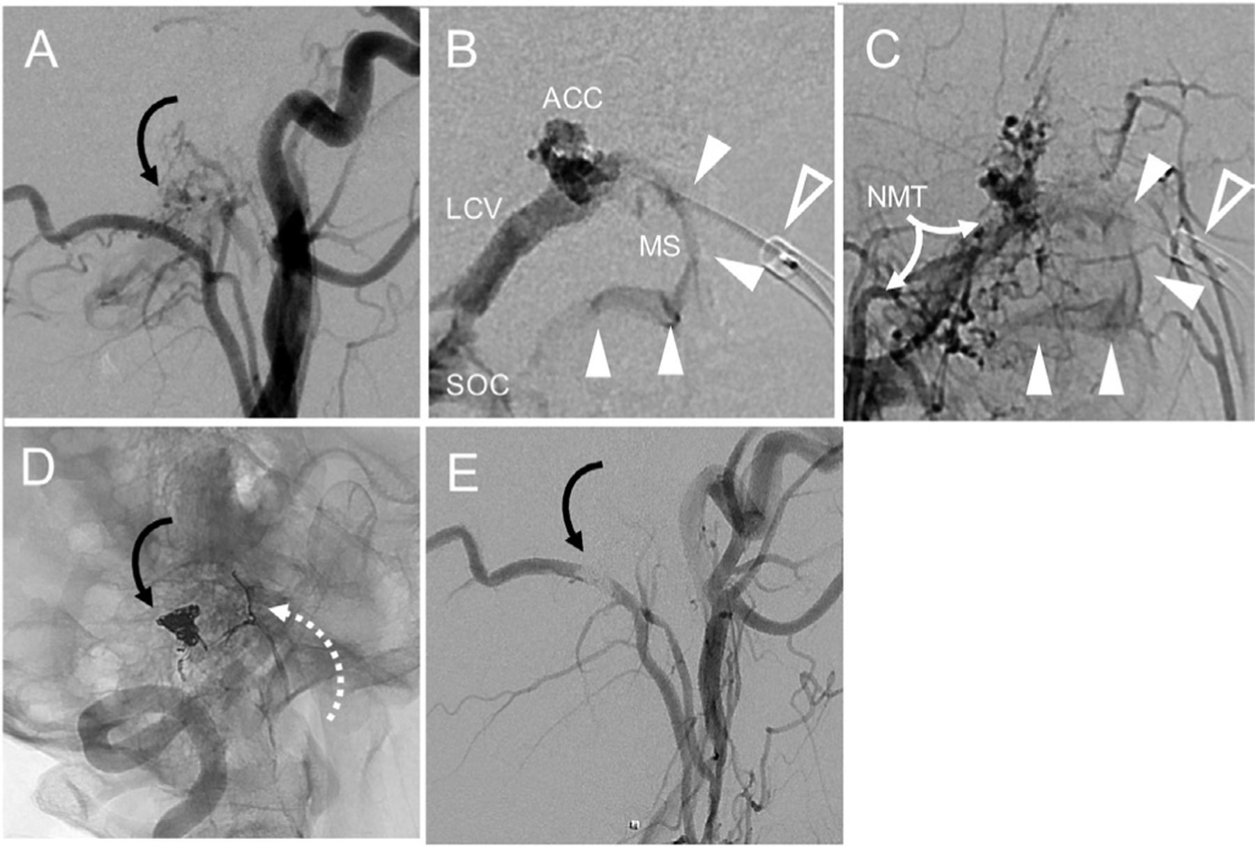
Figure 3.

A marginal sinus (MS) arteriovenous fistula shown (A, curved arrow), supplied by branches of the neuromeningeal trunk (NMT). Selective venography shows position of guide catheter (B, clear arrowhead) and microcatheter positioned near the fistula; white arrowheads show ‘wash-in’ from arteriovenous shunting. Selective ascending pharyngeal arteriography (C) shows NMT supply to fistula and confirms the position of the shunt (white arrowheads) from the venogram, facilitating targeted coil embolization. Postembolization unsubtracted image (D) shows configuration of coils in the MS region and evidence of unsuccessful prior Onyx embolization (dashed arrow) from another institution. Complete resolution of the shunt (black curved arrow) is shown on post-treatment angiogram (E). ACC, anterior condylar confluence; LCV, lateral condylar vein; SOC, Suboccipital cavernous sinus.
