
Fig. 1 |. Primary metastatic profiles of leukaemia metastasis.
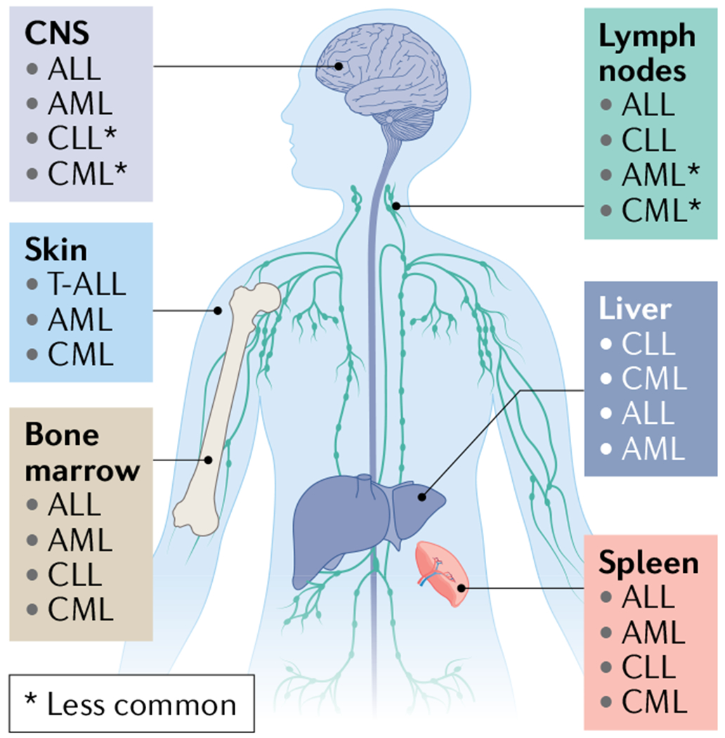
Acute lymphoblastic leukaemia (ALL) cells are typically initially found in the peripheral vascular system and the haematopoietic organs, including the bone marrow (BM)15, lymphatic system and spleen; however, central nervous system (CNS) involvement occurs in 5–10% of adult patients at diagnosis230 and can occur in up to 30–50% of patients in the absence of CNS-directed therapy. WHO classifies ALL and lymphoblastic lymphoma as a spectrum disorder and distinguishes these diseases according to clinical presentation favouring BM (involvement of >20% of blasts) or lymphatic tissue involvement, respectively15. Acute myeloid leukaemia (AML) primarily develops and spreads within the BM and less frequently involves other haematopoietic organs. It may invade non-haematopoietic organs including the gingiva, skin, muscle and CNS, although uncommonly. Chronic lymphocytic leukaemia/small lymphocytic lymphoma (CLL/SLL) is notable for its highly stereotyped clinical evolution: at the earliest stages, bloodstream lymphocytosis is the sole clinical finding, followed over time by the development of lymph node enlargement, then splenomegaly and in advanced stages, progressive BM involvement231. CLL and SLL are considered the same entity, but with differing disease presentation of bloodstream (CLL) or nodal involvement (SLL)232,233. Chronic myeloid leukaemia (CML) primarily involves the BM, peripheral circulation and spleen234. In rare cases including during evolution to blast crisis, CML expansion can result in infiltration into other less common sites, including the lymph nodes, liver, skin or CNS235–237. T-ALL, T cell ALL.