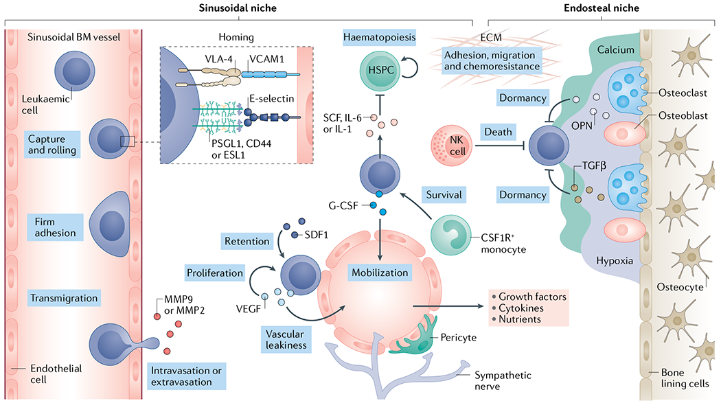
Fig. 2 |. The leukaemia bone marrow microenvironment.

Leukaemia cell homing is mediated by the chemokine stromal cell-derived factor 1 (SDF1)9,99–101 and adhesion molecules such as the integrin VLA-48,53–55 and E-selectin9 expressed by the vascular endothelium of fenestrated, sinusoidal bone marrow153 vessels. In this niche, growth factors, cytokines, extracellular matrix (ECM) components as well as stromal cells such as NG2+ cells, pericytes and colony stimulating factor 1 receptor-positive (CSF1R+) monocytes117 all serve to promote leukaemia cell growth and survival. As leukaemia cells colonize the bone marrow (BM), they deplete resident haematopoietic stem and progenitor cells (HSPCs) through the secretion of stem cell factor (SCF), creating a malignant niche that disrupts normal haematopoiesis123. Leukaemia cells may also migrate to pro-dormancy endosteal niches where osteopontin (OPN)35, transforming growth factor-β (TGFβ)238 and hypoxic conditions support a quiescent state, thus protecting them from chemotherapy. Niche factors such as granulocyte colony-stimulating factor (G-CSF) can mobilize leukaemia cells from the metastatic niche into circulation where they can seed distant sites201–203. Matrix-degrading enzymes such as elastase143, matrix metalloproteinase 2 (MMP2)92 and MMP9 (REFS90,239) can facilitate the extravasation process. Finally, immune cells such as natural killer (NK) cells may limit the survival of leukaemia cells in the BM240,241. PSGL1, P-selectin glycoprotein ligand 1; VCAM1, vascular cell adhesion molecule 1; VEGF, vascular endothelial growth factor.
